

What Is Retinal Imaging, and Why Does It Matter?
Retinal imaging gives your eye doctor a high-quality digital view of the back of your eye — the retina, macula, optic nerve, and blood vessels — often catching disease before you'd ever notice a symptom.
What Retinal Imaging Actually Shows
Retinal imaging captures a detailed picture of the structures at the back of your eye. It helps your provider identify and diagnose eye disease, including common conditions like glaucoma and macular degeneration, and shows the severity of an issue and how well it's responding to treatment over time.
Think of it as a window into the back of your eye — one that lets your provider see and monitor changes long before they'd show up as noticeable vision loss.
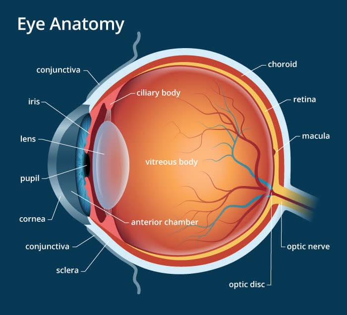
Anatomy of the Eye, Reviewed
Here's a simple breakdown of the structures your provider is actually looking at during an imaging exam.
The sensory membrane lining the inner back of the eye. It's home to rods and cones — your photoreceptors — which turn light into nervous signals sent to the brain's visual centers via the optic nerve.
A small, sensitive spot at the center of the retina responsible for central vision, visual acuity, and color vision — it's what lets you see fine detail while the rest of the retina handles peripheral vision.
A bundle of nerves at the back of the eye that acts as the eye's "messenger," carrying signals from the eye to the brain so it can register what you're seeing as an image.
The blood vessels of the eye and retina supply nutrients and remove waste — a healthy vascular network is essential to seeing clearly.
Types of Retinal Imaging
Not all retinal imaging looks the same. Your provider may use one or more of these techniques depending on what they need to see.
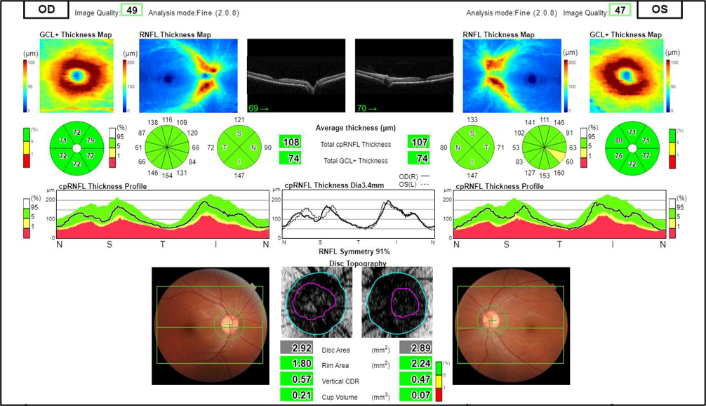
A non-invasive scan that uses light waves to capture cross-sections of the retina, giving a 3D look at eye structures and retinal thickness. You rest your chin on a support while the machine scans — results are instant, giving your provider immediate feedback.
A specialized camera with a low-power microscope captures a 30–50 degree color view of the retina and optic nerve. Your eyes are dilated first, and a bright light is used while a series of images of the back of the eye (the fundus) are taken.
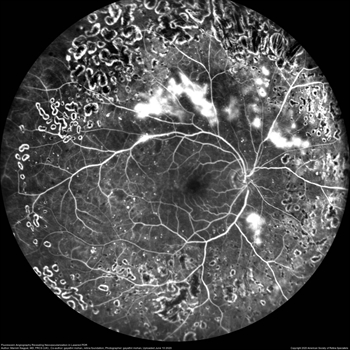
A technique used for decades: a dye is injected into the bloodstream and a specialized camera records blood flow within the retina after eye dilation. A series of bright lights and photos captures that flow, helping highlight focal lesions.
What to Expect
Depending on the imaging type, your provider may use dilating drops to widen your pupils for a clearer view of the back of your eye.
You'll sit in front of a specialized camera or scanner, usually resting your chin on a support to stay steady while the images are captured.
Many retinal imaging techniques, like OCT, produce results immediately — giving you and your provider instant feedback on the health of your eyes.
Retinal imaging can catch changes in your eyes long before you'd notice symptoms yourself. Talk to your eye doctor about whether retinal imaging should be part of your next exam.
Sources
- Advanced Retinal Imaging, American Society of Retina Specialists
- Retinal Imaging, Cleveland Clinic
- Retinal Imaging: Choosing the Right Method, American Academy of Ophthalmology
- Anatomy of the Eye, Eye Doctors of Brooklyn